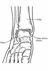
What is a Talar Dome Lesion?
 The ankle joint is composed of the bottom of the tibia (shin) bone and the top of the talus (ankle) bone. The top of the talus is dome-shaped and is completely covered with cartilage—a tough, rubbery tissue that enables the ankle to move smoothly. A talar dome lesion is an injury to the cartilage and underlying bone of the talus within the ankle joint. It is also called an osteochondral defect (OCD) or osteochondral lesion of the talus (OLT). “Osteo” means bone and “chondral” refers to cartilage.
Talar dome lesions are usually caused by an injury, such as an ankle sprain. If the cartilage doesn’t heal properly following the injury, it softens and begins to break off. Sometimes a broken piece of the damaged cartilage and bone will “float” in the ankle.
Signs and Symptoms
Unless the injury is extensive, it may take months, a year, or even longer for symptoms to develop. The signs and symptoms of a talar dome lesion may include:
- Chronic pain deep in the ankle—typically worse when bearing weight on the foot (especially during sports) and less when resting
- An occasional “clicking” or “catching” feeling in the ankle when walking
- A sensation of the ankle “locking” or “giving out”
- Episodes of swelling of the ankle—occurring when bearing weight and subsiding when at rest
Diagnosis
A talar dome lesion can be difficult to diagnose, because the precise site of the pain can be hard to pinpoint. To diagnose this injury, the foot and ankle surgeon will question the patient about recent or previous injury and will examine the foot and ankle, moving the ankle joint to help determine if there is pain, clicking, or limitation of motion within that joint.
Sometimes the surgeon will inject the joint with an anesthetic (pain-relieving medication) to see if the pain goes away for a while, indicating that the pain is coming from inside the joint.
X-rays are taken, and often an MRI or other advanced imaging tests are ordered to further evaluate the lesion and extent of the injury.
Treatment: Non-Surgical Approaches
Treatment depends on the severity of the talar dome lesion. If the lesion is stable (without loose pieces of cartilage or bone), one or more of the following non-surgical treatment options may be considered:
- Immobilization. Depending on the type of injury, the leg may be placed in a cast or cast boot to protect the talus. During this period of immobilization, non-weightbearing range-of-motion exercises may be recommended.
- Oral medications. Nonsteroidal anti-inflammatory drugs (NSAIDs), such as ibuprofen, may be helpful in reducing the pain and inflammation.
- Physical therapy. Range-of-motion and strengthening exercises are beneficial once the lesion is adequately healed. Physical therapy may also include techniques to reduce pain and swelling.
- Ankle brace. Wearing an ankle brace may help protect the patient from re-injury if the ankle is unstable.
When is Surgery Needed?
If non-surgical treatment fails to relieve the symptoms of talar dome lesions, surgery may be necessary. Surgery may involve removal of the loose bone and cartilage fragments within the joint and establishing an environment for healing. A variety of surgical techniques is available to accomplish this. The surgeon will select the best procedure based on the specific case.
Complications of Talar Dome Lesions
Depending on the amount of damage to the cartilage in the ankle joint, arthritis may develop in the joint, resulting in chronic pain, swelling and limited joint motion. Treatment for these complications is best directed by a foot and ankle surgeon, and may include one or more of the following
- Non-steroidal or steroidal anti-inflammatory medications
- Physical therapy
- Bracing
- Surgical intervention
Hours of Operation
Vancouver Foot And Ankle West
Monday:
8:00 am-4:00 pm
Tuesday:
Closed
Wednesday:
Closed
Thursday:
8:00 am-4:00 pm
Friday:
Closed
Saturday:
Closed
Sunday:
Closed
Vancouver Foot and Ankle East
Monday:
Closed
Tuesday:
Closed
Wednesday:
8:00 am-4:00 pm
Thursday:
8:00 am-4:00 pm
Friday:
8:00 am-4:00 pm
Saturday:
8:00 am-12:00pm (Alternating Saturday)
Sunday:
Closed
Battle Ground Foot and Ankle
Monday:
8:00 am-3:00 pm
(Alternating Monday)
Tuesday:
Closed
Wednesday:
8:00 am-4:00 pm
Thursday:
Closed
Friday:
Closed
Saturday:
Closed
Sunday:
Closed
Portland Foot and Ankle
Monday:
Closed
Tuesday:
8:00 am-3:00 pm
Wednesday:
Closed
Thursday:
Closed
Friday:
Closed
Saturday:
Closed
Sunday:
Closed
